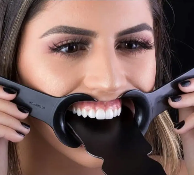
This is how we create beautiful smiles and help our patients achieve their goals.
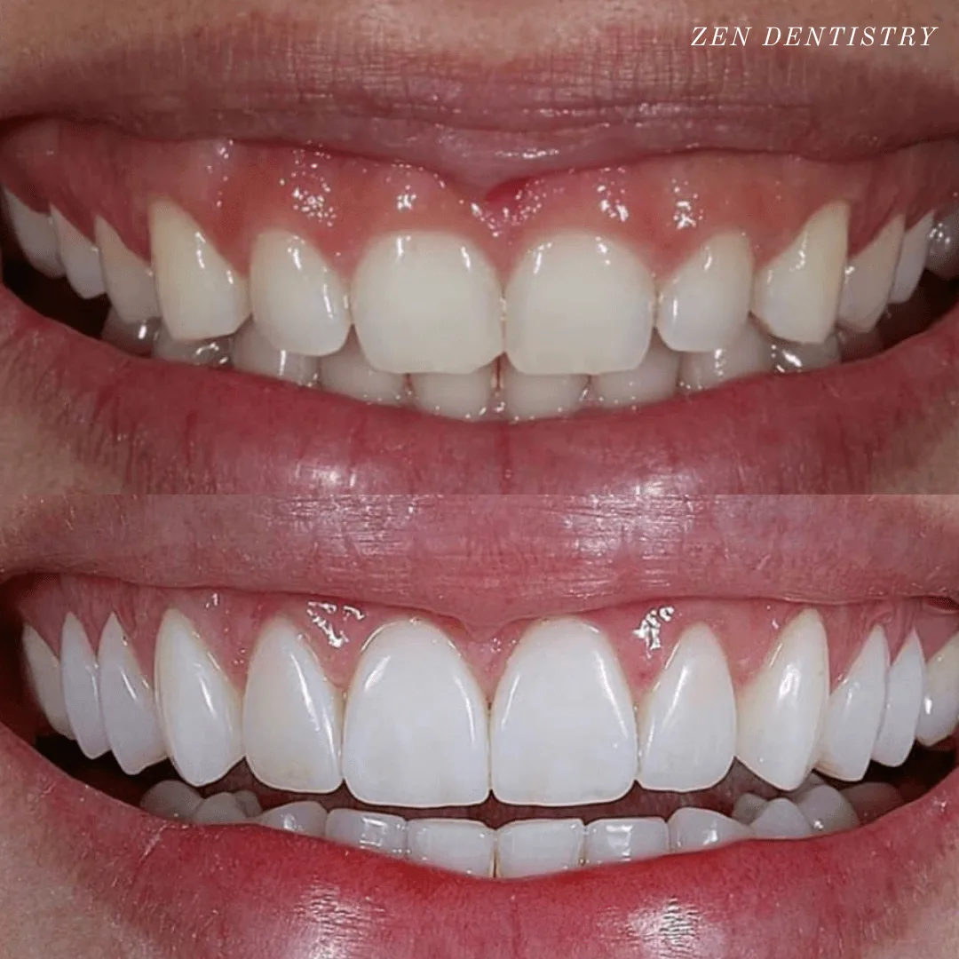
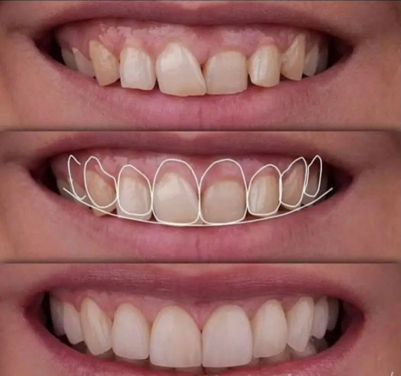
Patient presented to the office with a concern that the teeth were small and that too much gum shows when smiling. A comprehensive exam with x rays, pictures, smile analysis was done.
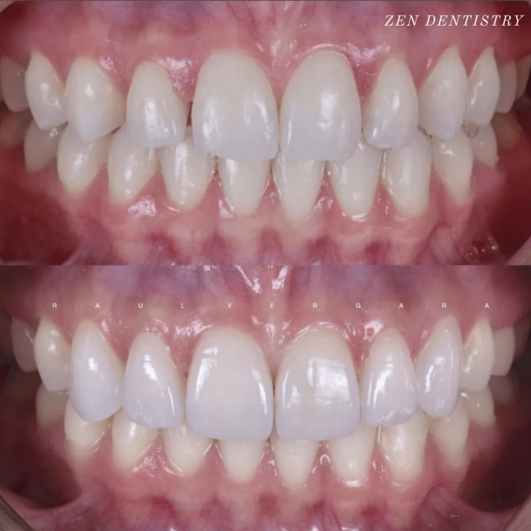
Patient presented to the office with a concern about spaces between the teeth and slight discoloration.
A detailed smile analysis with initial pictures, x rays, a scan of the teeth was used to fabricate a mock up smile.
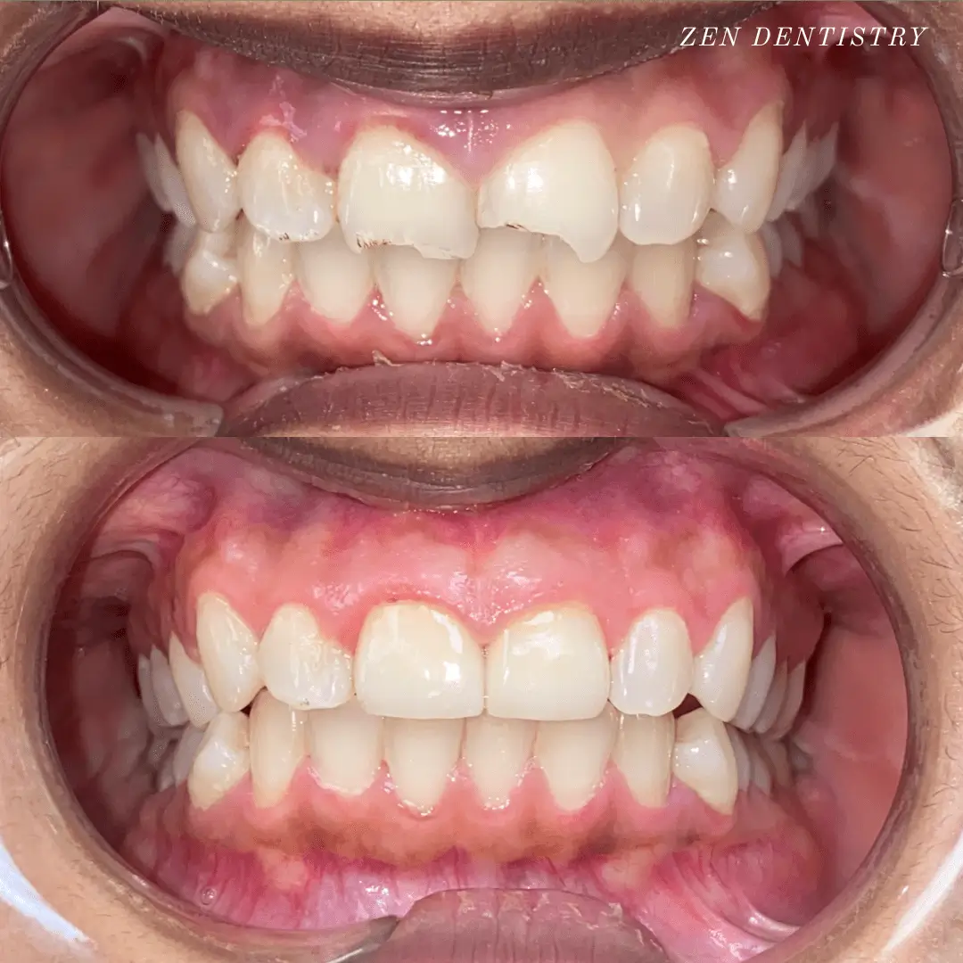
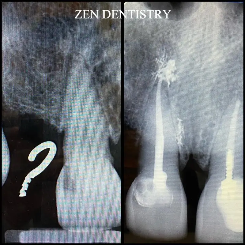
Patient presented to the office with trauma to front two teeth several weeks ago causing her severe pain.
After a thorough clinical exam which included endodontic screening and proper x rays led to the diagnosis of necrotic (dead) pulp tissue due to the force of trauma.
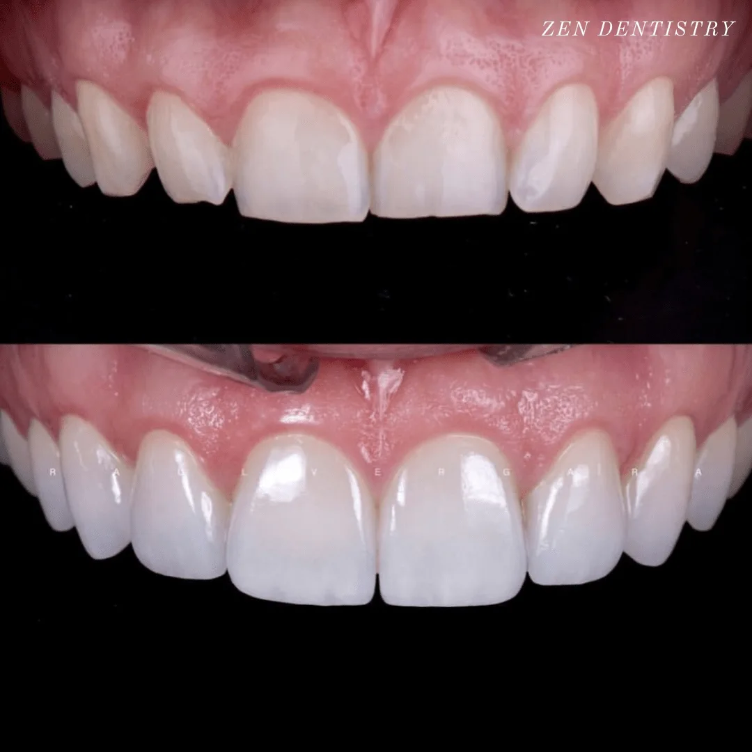
Patient presented to the clinic with the concern about the way smile looked.
A thorough aesthetic analysis was done and it was decided that the patient needed bigger teeth and gum contouring to compliment the facial anatomy.
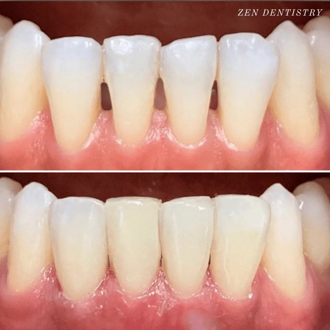
The patient presented to the office with a concern about spaces between lower front teeth.
After a detailed conversation with the patient and a thorough clinical exam, the patient decided to get composite bindings done to close the gaps.
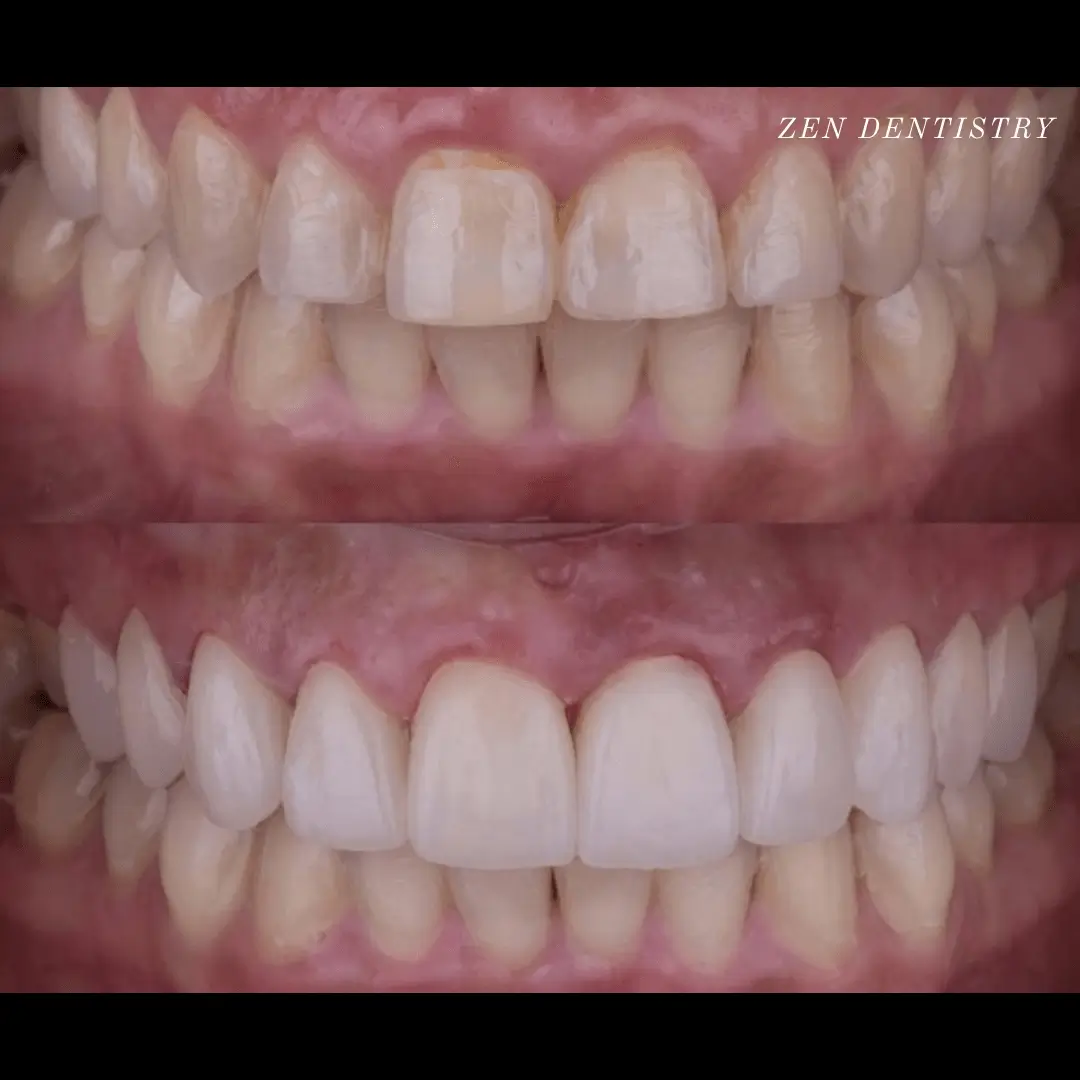
Patient presented to the
A thorough clinical exam, endodontic screening and proper x rays led to the diagnosis of irreversible pulpitis(inflamed pulp tissue) due to secondary dental caries with inflamed periodontal ligaments.
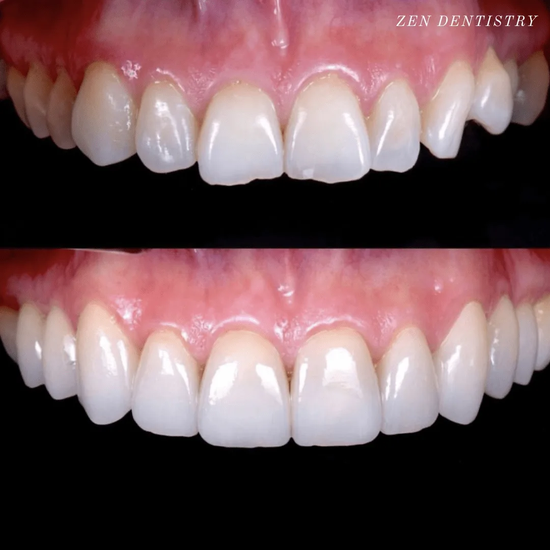
Patient presented to the
A thorough clinical exam, endodontic screening and proper x rays led to the diagnosis of irreversible pulpitis(inflamed pulp tissue) due to secondary dental caries with inflamed periodontal ligaments.
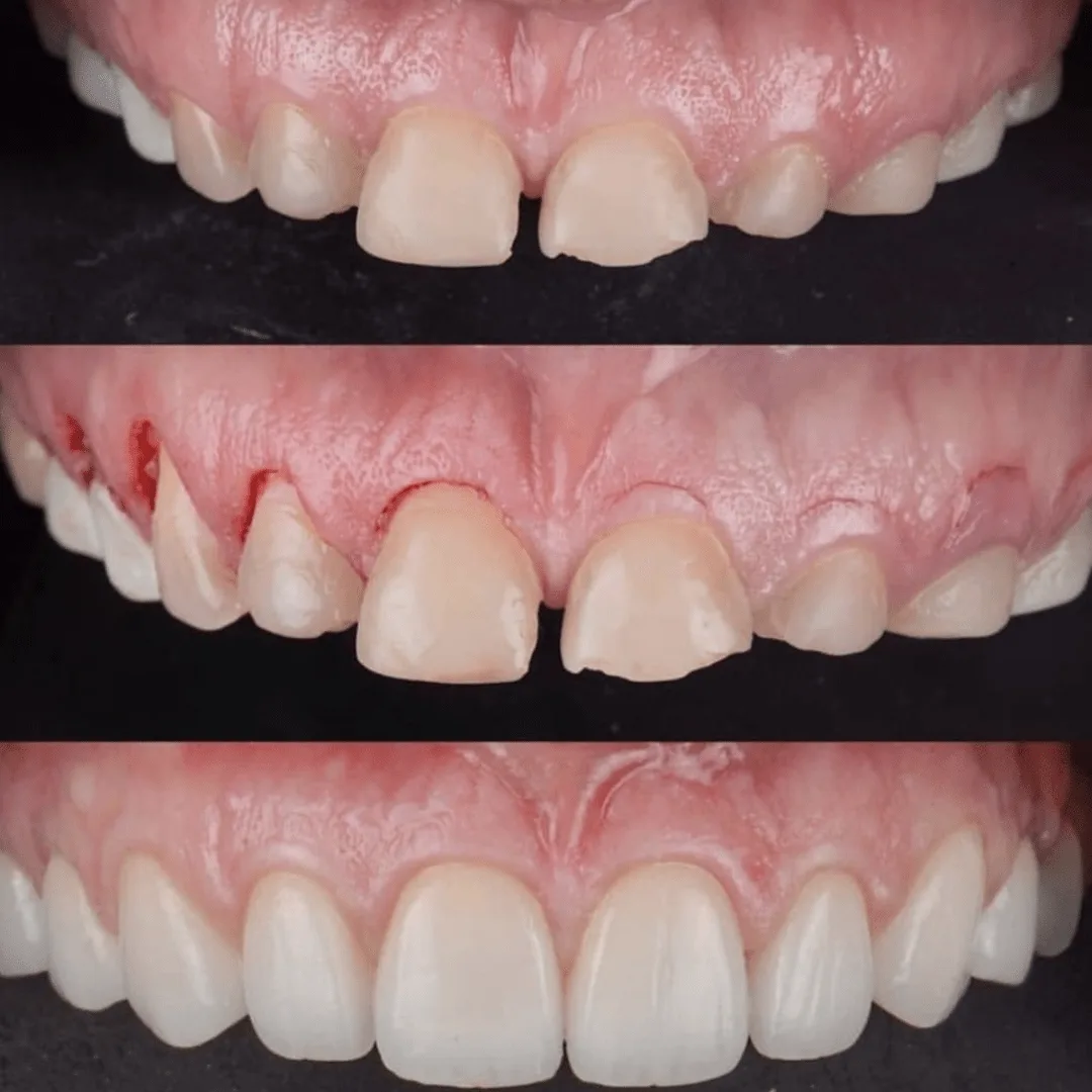
Patient presented to the office with a concern about the smile. Patient did not like the way teeth were and was causing severe emotional stress.
A detailed smile analysis was performed that included taking multiple pictures, x rays and scanning the teeth to create models to replicate the way patient bites.
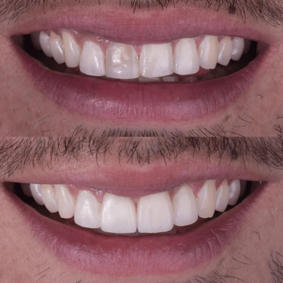
Patient presented to the office with a concern about front two teeth that had been discolored over time.
A detailed clinical exam and smile analysis was performed that included taking multiple pictures, x rays and scanning the teeth to create models to replicate the way patient bites.
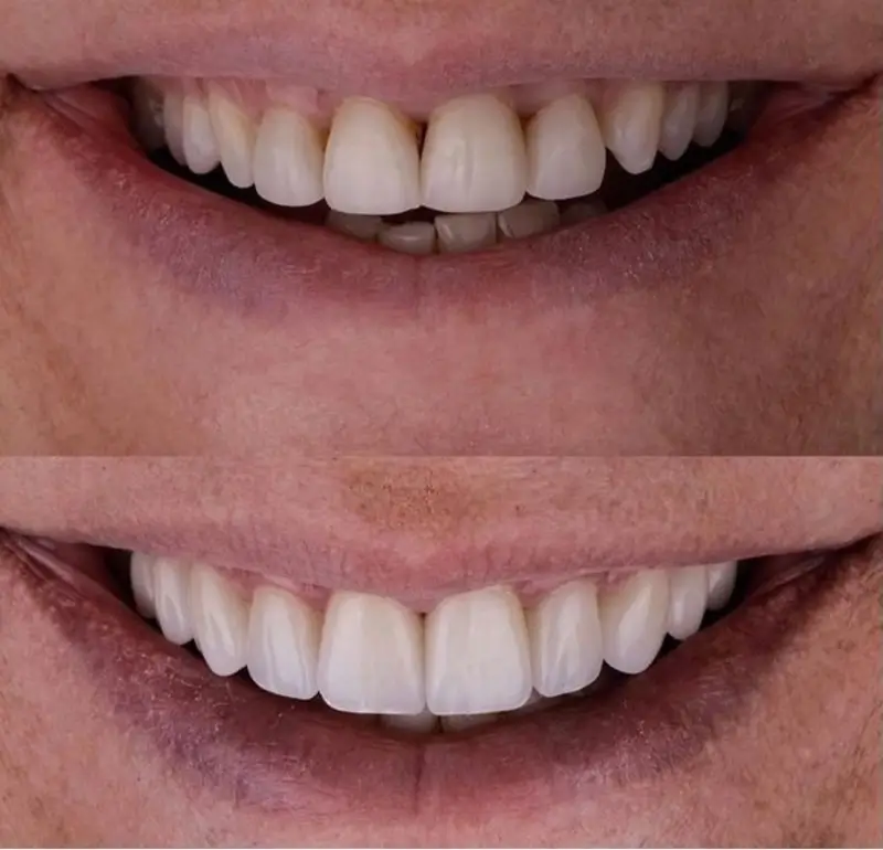
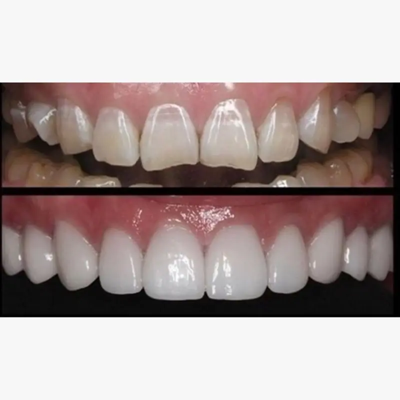
Patient presented to the office with a concern that the smile made them look old. A detailed clinical exam, smile analysis and understanding the patient expectations led us to a treatment plan that included 10 minimally prepped veneers to achieve a young and a balanced smile.
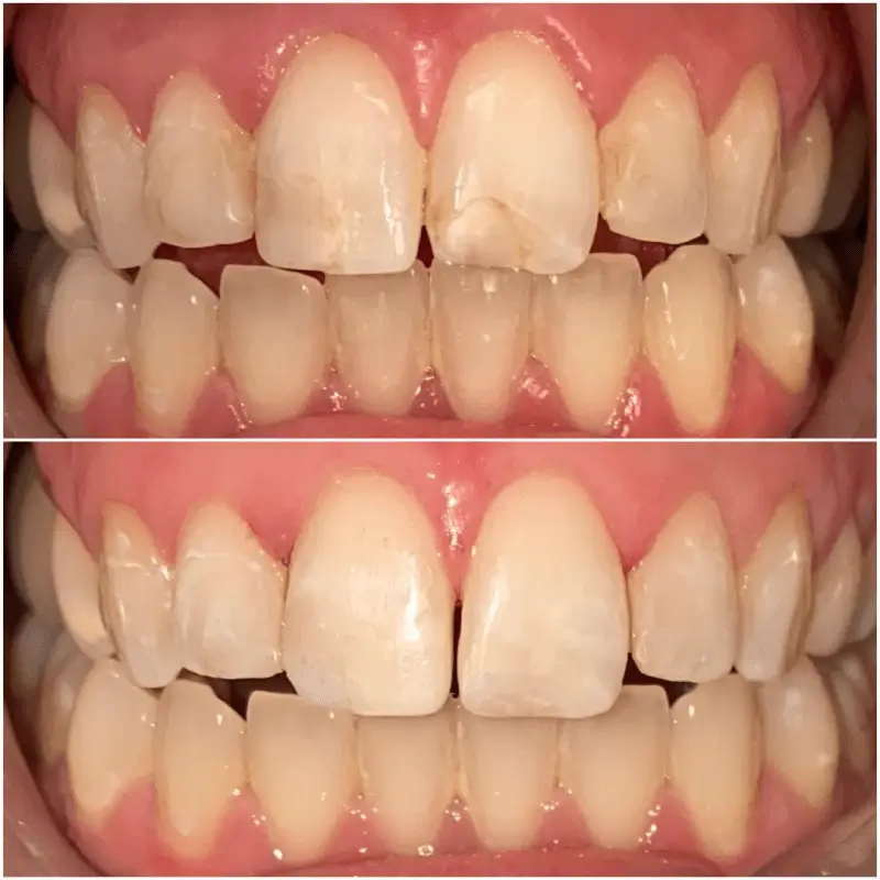
Patient presented to the office unhappy with the old composite restorations but did not want anything extensive. Patients goal was to re do the composite bondings.
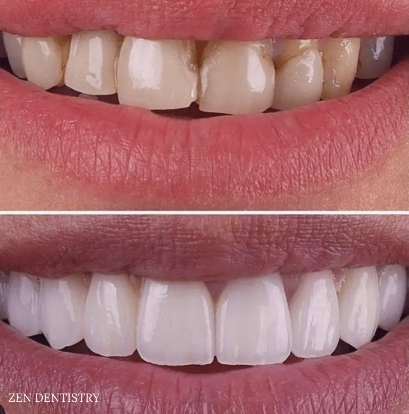
Patient presented to the office with the complaint of stained older restorations, minor crowding and alignment issues that had started to bother her. She did not want to undergo invisalign or traditional braces.
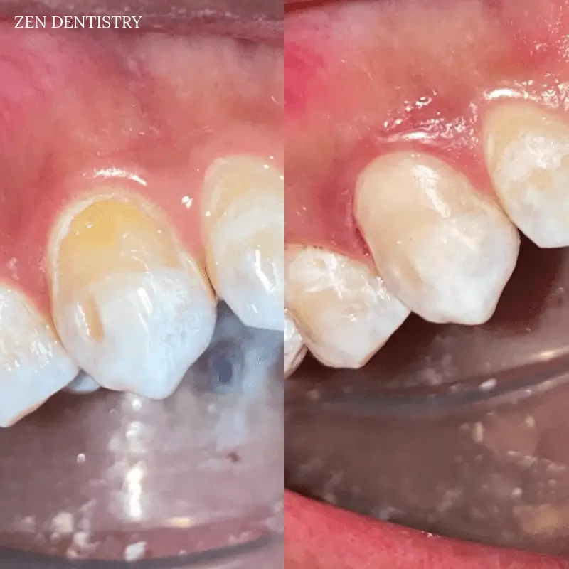
Patient presented to the office for bonding of this one tooth with a very specific request that we maintain the stains as best as we can to match the other teeth for time being.
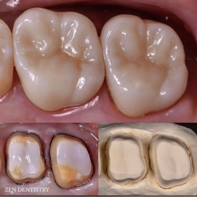
Patient presented to the office with a chief complaint of grinding his teeth at night. The patient’s molar teeth were quite significantly ground down from years of clenching that he needed root canals to be able to fix the issue of severe sensitivity.
Patient presented to the
A thorough clinical exam, endodontic screening and proper x rays led to the diagnosis of irreversible pulpitis(inflamed pulp tissue) due to secondary dental caries with inflamed periodontal ligaments.
Patient presented to the
A thorough clinical exam, endodontic screening and proper x rays led to the diagnosis of irreversible pulpitis(inflamed pulp tissue) due to secondary dental caries with inflamed periodontal ligaments.
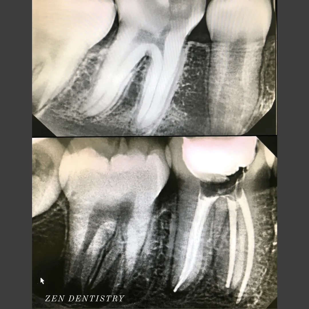
Patient presented to the
A thorough clinical exam, endodontic screening and proper x rays led to the diagnosis of irreversible pulpitis(inflamed pulp tissue) due to secondary dental caries with inflamed periodontal ligaments.
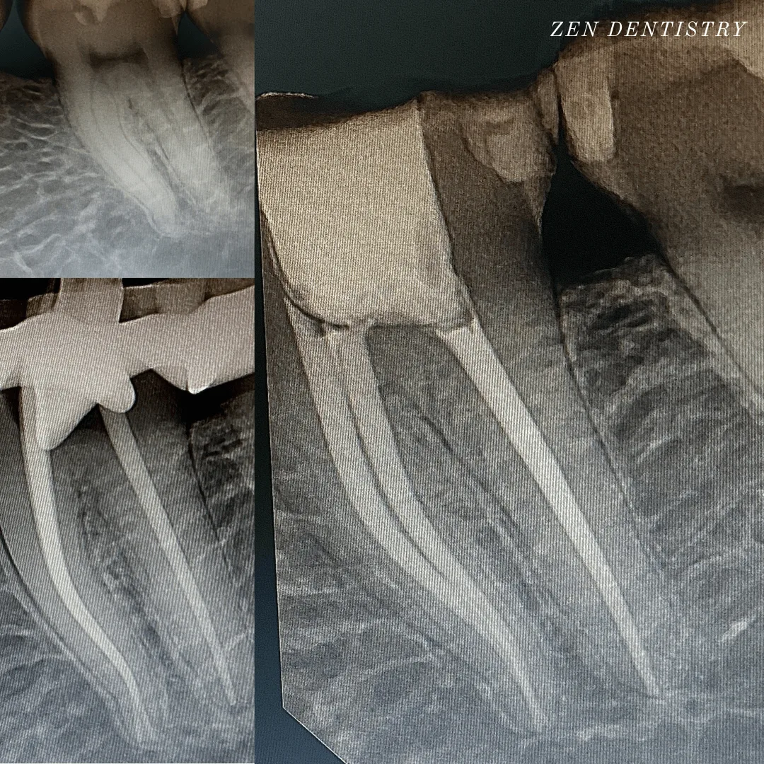
Patient presented to the office with a concern that the tooth has been causing severe constant pain that would become worse at night time and biting.
A detailed clinical exams including endodontic screening and x rays let to the diagnosis of pulp necrosis (dead nerve)due to gross dental caries that reached the nerve of the tooth with symptomatic apical periodontitis (inflamed ligaments).
Patient presented to the office with a concern that the tooth has been causing severe constant pain that would become worse at night time and biting.
A detailed clinical exams including endodontic screening and x rays let to the diagnosis of irreversible pulpitis (inflammation of the nerve) due to existing filling that was very close to the pulp and symptomatic apical periodontitis (inflamed ligaments).
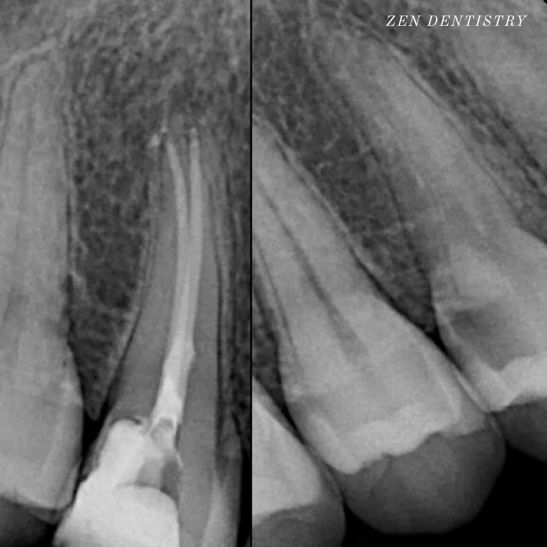
Patient presented to the office with a concern that the tooth has been causing severe constant pain that would become worse at night time.
A detailed clinical exams including endodontic screening and x rays let to the diagnosis of irreversible pulpitis (infected nerve) due to gross dental caries that reached the nerve of the tooth with asymptomatic apical periodontitis (normal ligaments).
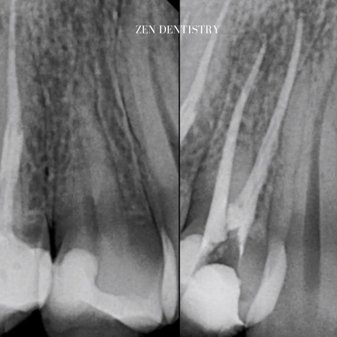
Patient presented with a complaint of severe constant pain that would exaggerate on laying down and biting.
Patient wanted to save the tooth.
A thorough clinical exam, endodontic screening and proper x rays led to the diagnosis of necrotic (dead) pulp due to dental caries with inflamed periodontal ligaments.
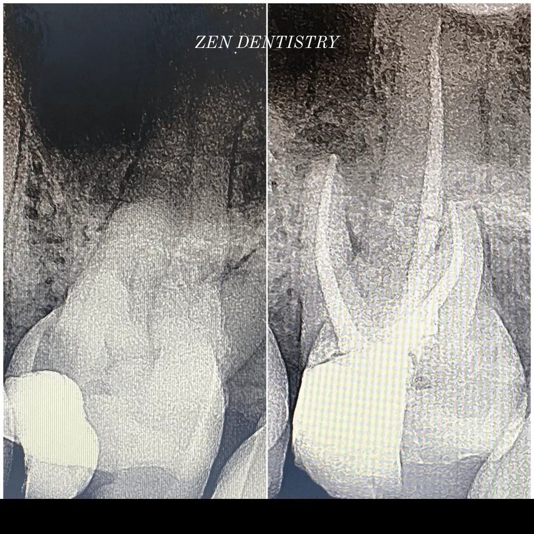
Patient presented with a complaint of severe constant pain that would exaggerate on laying down and biting.
A thorough clinical exam, endodontic screening and proper x rays led to the diagnosis of irreversible pulpitis(inflamed pulp tissue) due to secondary dental caries with inflamed periodontal ligaments.
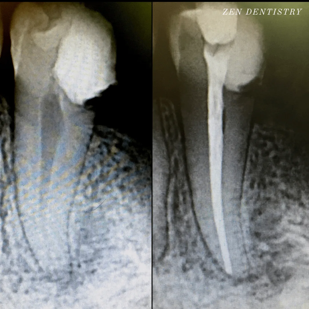
Patient presented with a complaint of severe constant pain that would exaggerate on laying down and biting.
A thorough clinical exam, endodontic screening and proper x rays led to the diagnosis of necrotic pulpal tissue(dead pulp tissue) due to secondary dental caries with inflamed periodontal ligaments.
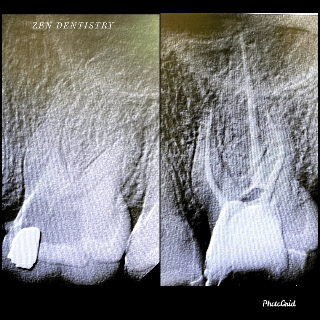
Patient presented to the office with a concern that the tooth had been hurting constantly since last few days.
The pain would become worse at night time and would wake the patient up from sleep.
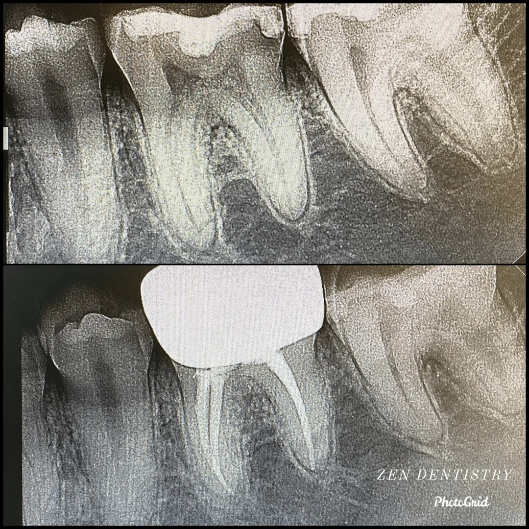
Patient presented with a complaint of severe constant pain that would exaggerate at night time and wake the patient up from sleep.
Patient wanted to save the tooth.
A thorough clinical exam, endodontic screening and proper x rays led to the diagnosis of irreversible pulpitis due to gross dental caries involving the pulp.
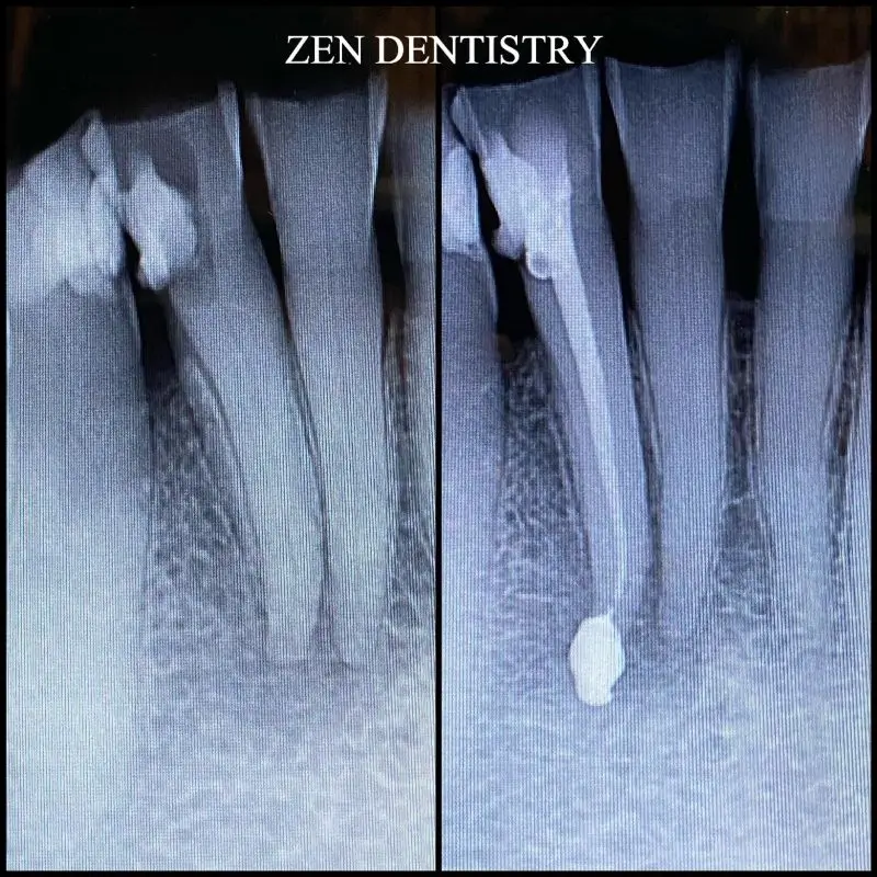
Patient presented to the office with a concern that the tooth has been causing severe constant pain that would become worse at night time and biting.
A detailed clinical exams including endodontic screening and x rays let to the diagnosis of pulp necrosis (dead nerve)due to gross dental caries that reached the nerve of the tooth with symptomatic apical periodontitis (inflamed ligaments).

